
1 / 7
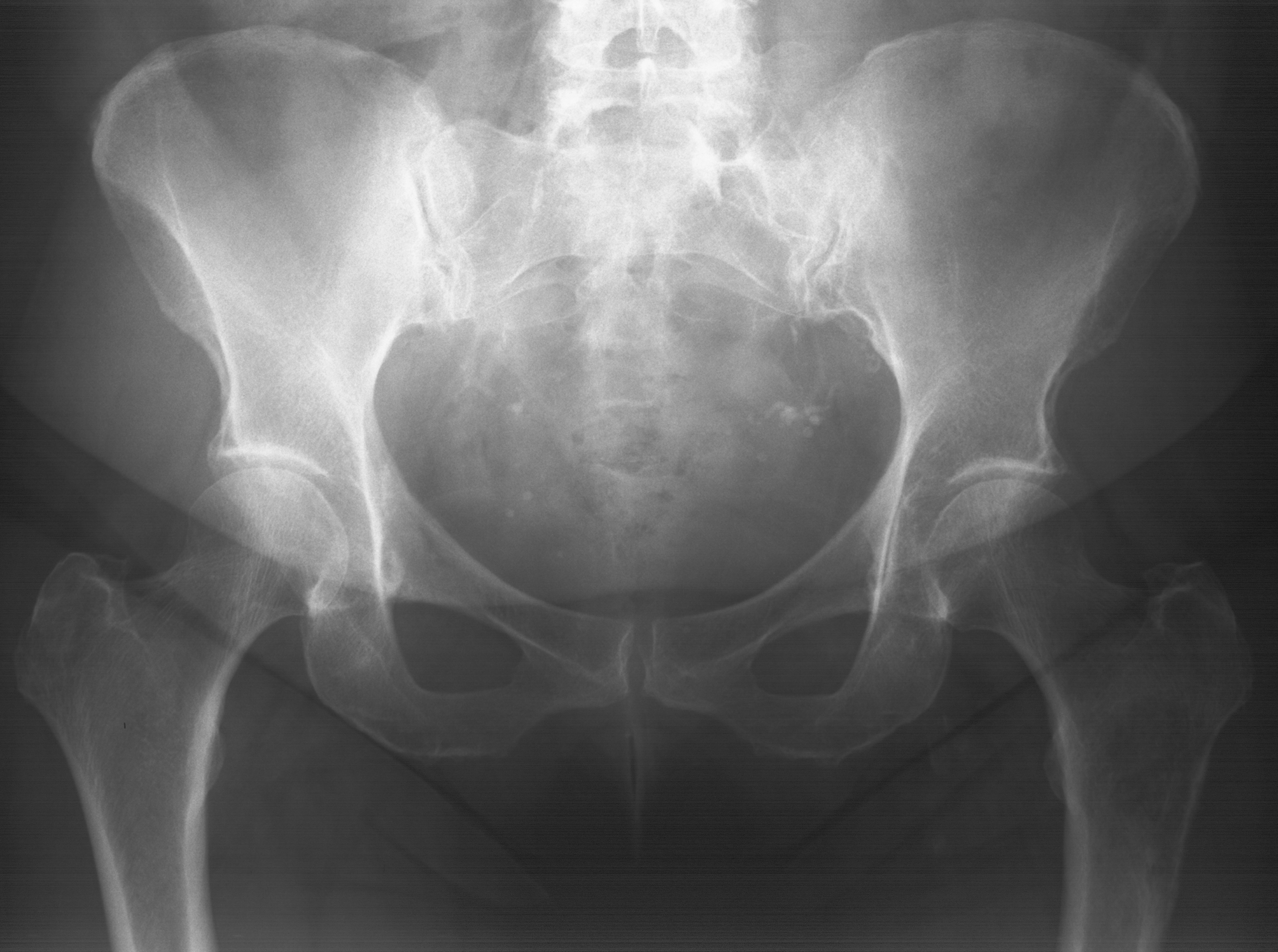
1. AP pelvis
HipBasic
Patient66y Female
Case ID#1836
ModalitiesX-ray, MRI
48 views
3 July 2026
Tags
Case 1836
Clinical History
66-year-old female presenting with hip pain for 8 weeks. Initial radiograph of the pelvis interpreted as normal. Further imaging was performed 2 months later due to progressive symptoms.