
1 / 7
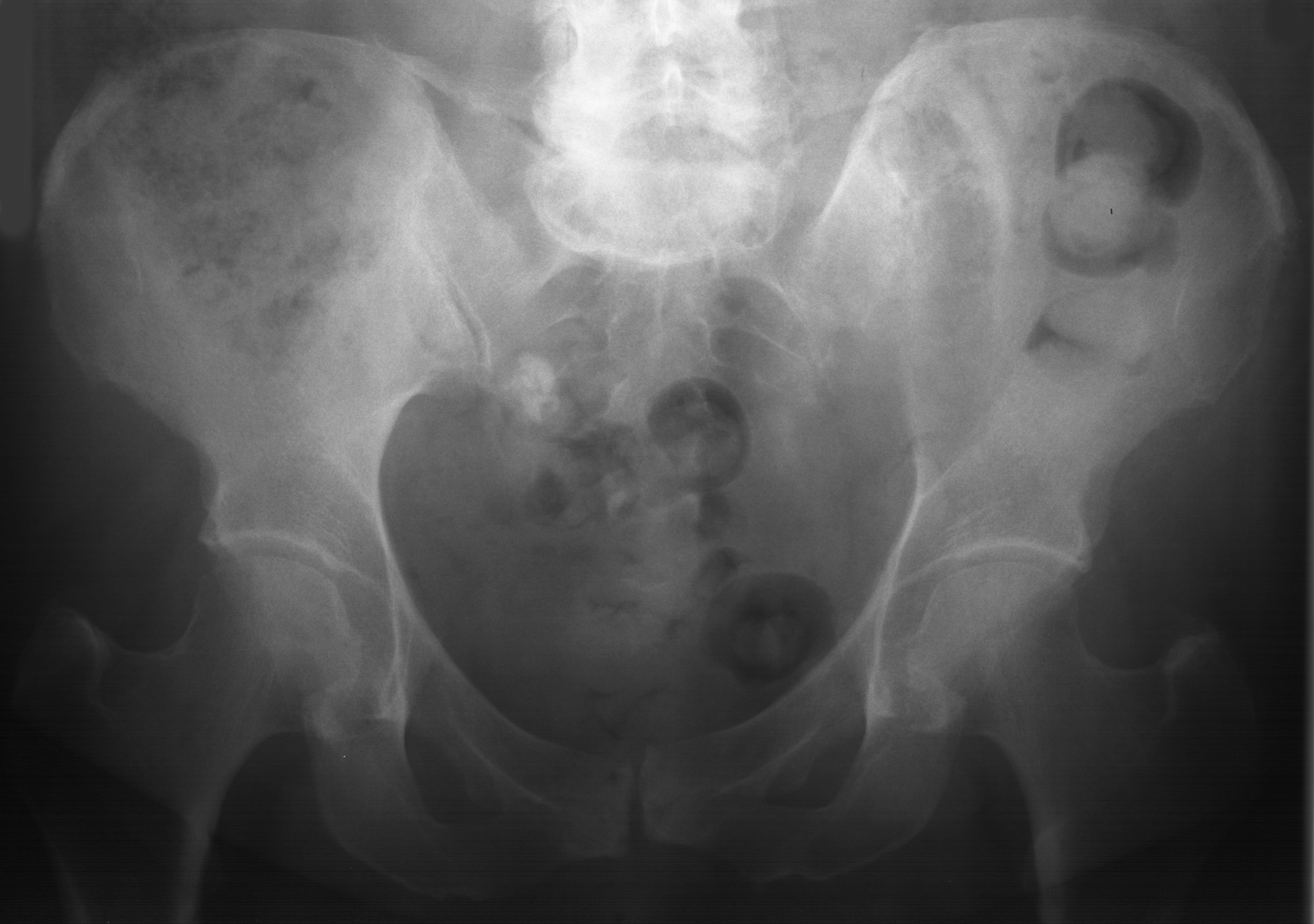
1. AP pelvis
HipBasic
Patient59y Female
Case ID#1818
ModalitiesX-ray, Nuclear Medicine, CT, MRI
37 views
3 July 2026
Tags
Case 1818
Clinical History
59-year-old female presenting with low back and hip pain. Radiograph at presentation reported as normal. Further imaging performed 26 months later. CT chest, abdomen and pelvis noted to be normal apart from the iliac lesion. Thyroidectomy for 'benign' disease 16 years ago.